The Role of Ultrasound in Diagnosing Trigger Finger
Trigger finger, a condition medically known as stenosing tenosynovitis, affects the tendons in the fingers or thumb, causing them to lock or catch during movement. While this condition is common and often painful, accurate diagnosis is crucial for effective treatment. Among various diagnostic tools, ultrasound has emerged as a game-changer, offering precise, non-invasive insights into the structures of the affected finger. This article explores how ultrasound plays a pivotal role in diagnosing the trigger finger and why it is becoming the preferred method for healthcare providers.
Understanding Trigger Finger
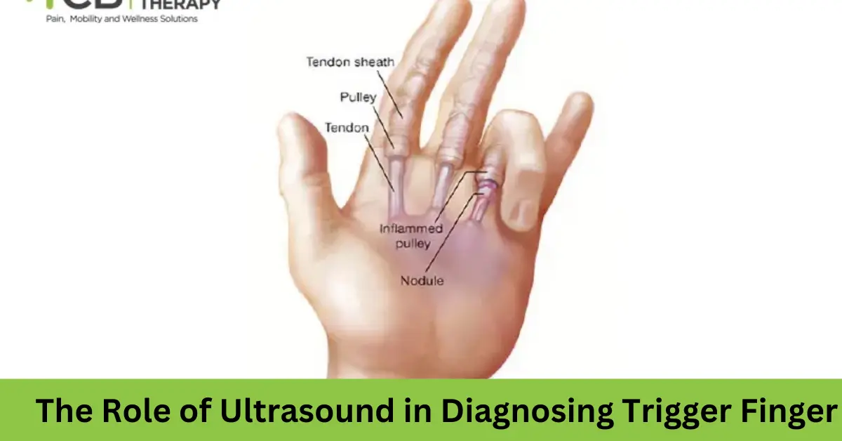
Trigger finger occurs when the tendons responsible for bending the fingers become inflamed or irritated. Tendons are surrounded by a protective sheath, and in the trigger finger, this sheath narrows, restricting smooth tendon movement. The result? A painful snapping or locking sensation as the finger moves.
The condition is most common among individuals engaged in repetitive hand activities, such as typing or gripping tools. It’s also more prevalent in people with underlying health issues like diabetes, rheumatoid arthritis, or carpal tunnel syndrome. Trigger fingers can range from mild discomfort to severe cases where the finger becomes stuck in a bent or extended position.
What is Ultrasound?
Ultrasound, also called sonography, is a diagnostic tool that uses high-frequency sound waves to create real-time images of tissues inside the body. It’s non-invasive, painless, and does not involve radiation, making it a safe option for diagnosing a wide range of conditions.
For musculoskeletal disorders like the trigger finger, ultrasound can visualize tendons, sheaths, and surrounding tissues, providing clear and detailed images that aid in diagnosis and treatment planning.
The Role of Ultrasound in Diagnosing Trigger Finger
Visualizing Tendon Movement
One of the biggest advantages of ultrasound is its ability to capture live, dynamic images of tendon movement. For the trigger finger, this means seeing exactly how the affected tendon behaves as the finger moves, helping to confirm the presence of abnormalities such as thickened tendons or narrowed sheaths.
Detecting Inflammation and Swelling
Ultrasound can reveal signs of inflammation, including swelling or fluid accumulation around the tendon sheath. This is particularly useful in the early stages of the trigger finger, where symptoms might not be severe but treatment could prevent further progression.
Measuring Tendon Thickness
Studies show that increased tendon thickness is a hallmark of the trigger finger. Ultrasound provides precise measurements of tendon size, allowing healthcare providers to identify whether the tendon is abnormally thickened—a critical diagnostic criterion.
Differentiating from Other Conditions
Trigger finger symptoms can overlap with other hand conditions, such as arthritis or tendon injuries. Ultrasound can help distinguish between these by offering a detailed view of the affected structures. For instance, it can identify bony abnormalities or rule out tears in the tendons.
Assessing Severity
Ultrasound not only confirms the diagnosis but also helps evaluate the severity of the condition. By identifying how much the tendon sheath is narrowed and how restricted the tendon movement is, clinicians can tailor treatment plans to the individual’s needs.
Benefits of Ultrasound in Trigger Finger Diagnosis
- Non-Invasive and Painless
Unlike other imaging techniques like MRI, ultrasound does not require injections, radiation, or confined spaces, making it a patient-friendly option. - Real-Time Imaging
Ultrasound captures the tendon in motion, providing dynamic insights that static imaging methods cannot. This is particularly important for trigger finger, where movement often reveals the problem. - Cost-Effective
Compared to MRI or other advanced imaging tools, ultrasound is more affordable, reducing the financial burden on patients while still offering accurate results. - Accessibility
Ultrasound machines are widely available in hospitals and clinics, making it easier for patients to access this diagnostic tool without long waiting periods.
Case Study: Ultrasound in Action
Imagine a 45-year-old patient presenting with pain and a locking sensation in their middle finger. A physical exam suggests a trigger finger, but the symptoms are mild, and the doctor wants to confirm the diagnosis before proceeding with treatment. An ultrasound scan is performed, revealing a thickened flexor tendon and swelling within the tendon sheath. Dynamic imaging shows restricted tendon movement, especially during finger flexion. With these clear findings, the doctor confirms the diagnosis and initiates appropriate treatment, such as corticosteroid injections or physical therapy, avoiding unnecessary delays.
Advancing Treatment Through Ultrasound
Beyond diagnosis, ultrasound can also guide treatments for the trigger finger. For instance:
- Guided Corticosteroid Injections: Ultrasound ensures that injections are precisely delivered to the affected tendon sheath, improving efficacy and reducing the risk of complications.
- Post-Treatment Monitoring: Ultrasound can track progress after treatments, such as physical therapy or surgery, ensuring that the tendon is healing properly.
Challenges and Limitations
While ultrasound offers numerous advantages, it’s not without limitations. Its effectiveness depends heavily on the skill and experience of the operator. Poor technique can lead to misinterpretation of images, potentially delaying or complicating treatment. Additionally, while ultrasound is excellent for soft tissue imaging, it may not provide detailed views of bone structures.
Conclusion
Ultrasound has revolutionized the way the trigger finger is diagnosed and managed. Its ability to provide real-time, detailed images of tendon movement and surrounding tissues makes it an invaluable tool for healthcare providers. Non-invasive, cost-effective, and widely accessible, ultrasound is bridging the gap between early diagnosis and effective treatment.
